医院检查报告单中译英翻译,加拿大律师公证
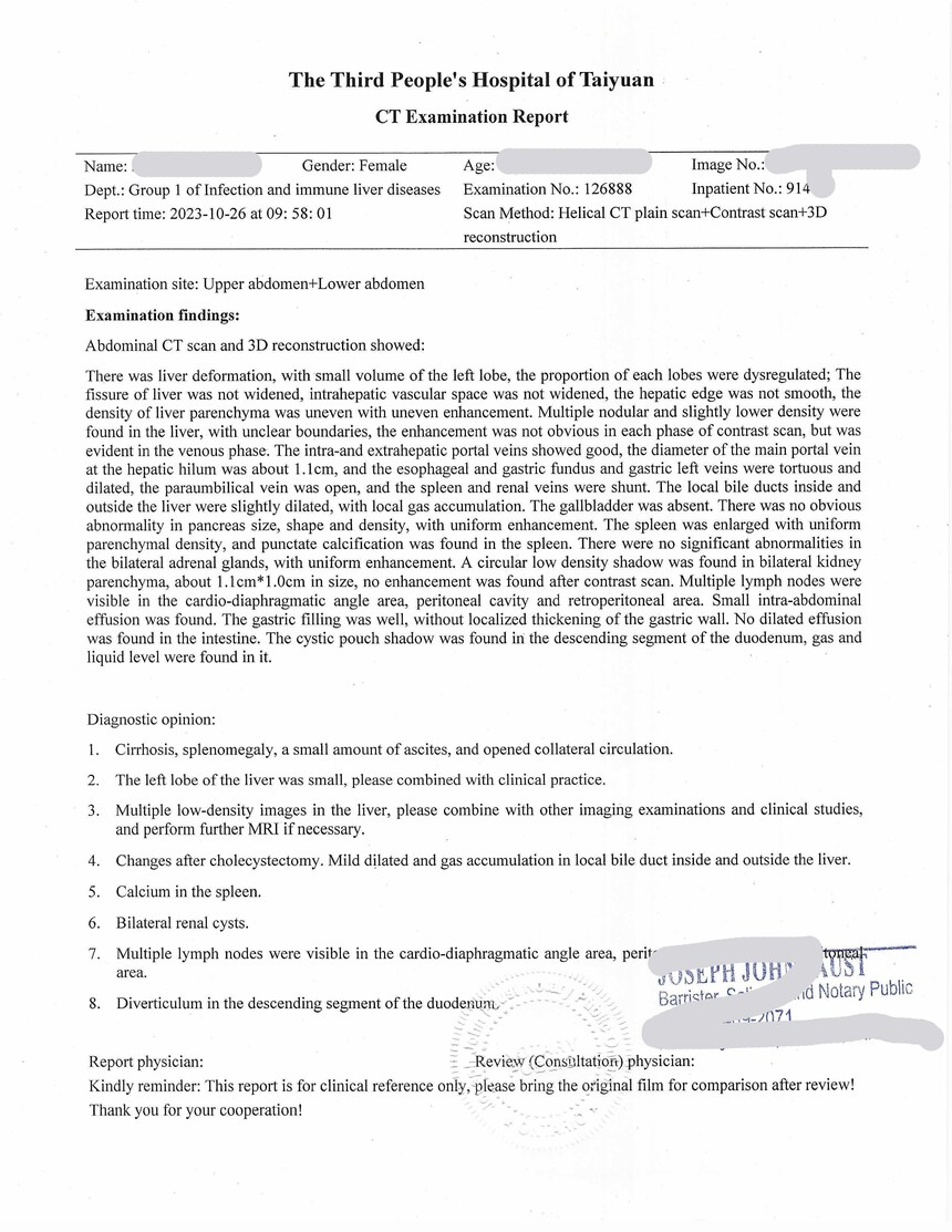
The Third People's Hospital of Taiyuan
CT Examination Report
Name: XXXX Gender: Female Age: 66y Image No.: XXXX
Dept.: Group 1 of Infection and immune liver diseases Examination No.: XXXX Inpatient No.: XXXX
Report time: 2023-10-26 at 09: 58: 01 Scan Method: Helical CT plain scan+Contrast scan+3D reconstruction
Examination site: Upper abdomen+Lower abdomen
Examination findings:
Abdominal CT scan and 3D reconstruction showed:
There was liver deformation, with small volume of the left lobe, the proportion of each lobes were dysregulated; The fissure of liver was not widened, intrahepatic vascular space was not widened, the hepatic edge was not smooth, the density of liver parenchyma was uneven with uneven enhancement. Multiple nodular and slightly lower density were found in the liver, with unclear boundaries, the enhancement was not obvious in each phase of contrast scan, but was evident in the venous phase. The intra-and extrahepatic portal veins showed good, the diameter of the main portal vein at the hepatic hilum was about 1.1cm, and the esophageal and gastric fundus and gastric left veins were tortuous and dilated, the paraumbilical vein was open, and the spleen and renal veins were shunt. The local bile ducts inside and outside the liver were slightly dilated, with local gas accumulation. The gallbladder was absent. There was no obvious abnormality in pancreas size, shape and density, with uniform enhancement. The spleen was enlarged with uniform parenchymal density, and punctate calcification was found in the spleen.There were no significant abnormalities in the bilateral adrenal glands, with uniform enhancement. A circular low density shadow was found in bilateral kidney parenchyma, about 1.1cm*1.0cm in size, no enhancement was found after contrast scan. Multiple lymph nodes were visible in the cardio-diaphragmatic angle area, peritoneal cavity and retroperitoneal area. Small intra-abdominal effusion was found. The gastric filling was well, without localized thickening of the gastric wall. No dilated effusion was found in the intestine. The cystic pouch shadow was found in the descending segment of the duodenum, gas and liquid level were found in it.
Diagnostic opinion:
1. Cirrhosis, splenomegaly, a small amount of ascites, and opened collateral circulation.
2. The left lobe of the liver was small, please combined with clinical practice.
3. Multiple low-density images in the liver, please combine with other imaging examinations and clinical studies, and perform further MRI if necessary.
4. Changes after cholecystectomy. Mild dilated and gas accumulation in local bile duct inside and outside the liver.
5. Calcium in the spleen.
6. Bilateral renal cysts.
7. Multiple lymph nodes were visible in the cardio-diaphragmatic angle area, peritoneal cavity and retroperitoneal area.
8. Diverticulum in the descending segment of the duodenum.
Report physician: Review (Consultation) physician:
Kindly reminder: This report is for clinical reference only, please bring the original film for comparison after review! Thank you for your cooperation!
The Third People's Hospital of Taiyuan
Ultrasound examination reportName: XXXX Gender: Female Age: 66y
Examination No.: XXXXX Inpatient No.: XXXX Ultrasound No.: XXXX
Application Dept.: Group 1 of Infection and immune liver diseases Examination equipment: PHILIPS EPIQ5 Examination site: Liver, gallbladder, pancreas, spleen, kidney, portal vein, ascites positioning, pleural fluid positioning
Examination parameters:
Left lobe thickness of liver 2.5cm Left lobe length of liver 3.3cm Maximum slope of the right lobe 13.2cm Long diameter of spleen 13.9cm
Intercostal thick diameter of the spleen 5.8cm Subcostal diameter 3.5cm Pancreatic head 1.1cm Body 0.8cm
Tail 1.1cm Left kidney 8.9*4.4cm Right kidney 9.8*3.2cm Main portal vein width 0.8cm
Blood flow velocity of the main portal vein 129cm/s Common bile duct width 0.4cm
Examination findings:
A massive intestinal tympanites reflex was found in the abdominal cavity.
The left lobe of the liver was small, the capsule of the right lobe of the liver was undulating, rough echo and uneven distribution within the liver parenchymal were found, multiple small hypoechoic reflected areas were found in the liver parenchyma with unclear boundaries and without blood flow signal; The right lobe of the liver could found a small strong echo, with acoustic shadow, the size was 0.5cm, there was no expansion of the tube around it; A heterogeneous echo region was visible in the right lobe of the liver, with unclear borders and the range was about 3.1*2.2cm, with obvious abnormal blood flow signal; Portal vein, splenic vein, and superior mesenteric vein all blood flow to the liver. In the left branch sac of the portal vein, a pipe structure extended to the liver surface, the inner diameter was 0.6cm, within which the venous spectrum from the liver could be found, Vmax: 17.2cm/s.
After cholecystectomy, there was no expansion of the interhepatic and extrahepatic bile ducts.
There was no abnormal echo of pancreas size and shape, and no dilatation of the main pancreatic duct.
A cystic dark region was visible in the middle pole of the left kidney with a smooth wall, and an enhancement effect in the posterior wall was ? cm, there was no abnormality in the remaining structures. The right kidney was normal in size and showed no structural abnormalities.
The long diameter of spleen: 13.9cm.
The liquid dark zone 0.9cm in size was visible above the bladder.
There was no free liquid dark zone in the bilateral thorax.
Examination suggestions:
1. Cirrhosis, it might be multiple small nodules in the liver; Calcification lesions in the right lobe of the liver; Heterogenous echo of the right lobe of the liver, and its nature? The paralignal vein was opened.
2. Splenomegaly
3. Left renal cyst
4. No abnormalities were observed in pancreas and right kidney
5. Ascites; No pleural effusion was found.
Regular review is recommended
Examination time: 2023-10-21 Examined physician: XXXX
Inputted by: XXXX
Ultrasound examination is affected by many factors in the patient himself . (obesity, gas disturbance, poor coordination, special lesion location, different stages of the disease, etc.) This report is for clinical reference only and is valid for this examination only.
第一页:律师宣誓页




20242072
推荐
-

-

QQ空间
-

新浪微博
-

人人网
-

豆瓣